(626) 323-9443
 432 W Foothill Blvd, Glendora, CA 91741, USA

In pain? Call (626) 323-9443 for same-day care. In pain right now? Call (626) 323-9443 — same-day emergency appointments available.
In pain? Call (626) 323-9443 for same-day care. In pain right now? Call (626) 323-9443 — same-day emergency appointments available.
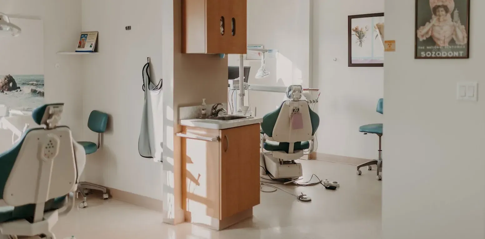
Clear digital imaging that reveals what a visual exam alone can't, helping us catch problems while they're still small and simple to treat.
A lot of dental trouble starts where your dentist's eyes can't reach: between teeth, below the gumline, inside the root, or deep within the jawbone. Cavities can form quietly between tight contact points for months before they ever cause a twinge of pain. Bone loss from gum disease can progress steadily without any visible warning on the surface. By the time a problem becomes obvious to the naked eye, it has often already grown more complicated and more costly to treat.
That's exactly why dental X-rays are a routine part of care at Glendora Healthy Smiles. Our digital imaging system lets Dr. and our clinical team see the full picture of your oral health, not just the parts visible during a standard exam. We use this information to catch decay between teeth, evaluate bone density around your roots, check the position of unerupted or impacted teeth, and confirm whether an old filling or crown is still sealing properly.
We know some patients worry about radiation exposure or discomfort from X-rays. Our digital sensors use significantly less radiation than older film-based systems, and the process itself takes just a few minutes. We'll always explain why we're recommending an image, what we're looking for, and how it factors into your treatment plan, so you're never left guessing.

Good imaging does far more than confirm a diagnosis you already suspect. It changes the entire direction of your treatment plan, often sparing you from larger procedures down the road and giving your dental team the confidence to recommend exactly the right course of action.
X-rays reveal decay forming between teeth long before it would ever show up on a visual exam or cause sensitivity. Treating a small cavity now, with a simple filling, is far less invasive and far less expensive than addressing deep decay that has reached the nerve.
Imaging shows the bone level surrounding each tooth root, which is how we track gum disease progression over time. Early detection of bone loss allows us to intervene with periodontal care before a tooth becomes loose or unstable.
An abscess or infection at the tip of a tooth root often causes no visible signs until it has grown substantially. X-rays detect dark shadows at the root tip that signal infection, letting us start treatment before you experience a painful flare-up.
Panoramic images show the full arrangement of teeth, roots, and jaw structure, including teeth that haven't erupted yet. This information is essential for planning braces, Invisalign, or extractions with precision and confidence.
We use imaging to check that old crowns, fillings, and root canals are still sealed properly and haven't developed gaps or decay underneath. Catching a failing restoration early prevents the need for a more extensive and costly replacement later.
X-rays reveal the exact angle and position of wisdom teeth beneath the gumline, which tells us whether they're likely to cause crowding, pain, or infection. We can plan removal before symptoms ever start.
Our digital sensors produce a usable image in seconds and use a fraction of the radiation of traditional film X-rays. You get accurate diagnostic information without a long wait or unnecessary exposure.
Not every situation that calls for an X-ray comes with obvious pain. Many issues that imaging uncovers are completely silent in their early stages, which is exactly why we recommend keeping up with the following warning signs and routine imaging intervals.
Ongoing sensitivity to hot, cold, or sweets can point to decay that has progressed closer to the nerve, even if it isn't visible on the surface.
Persistent pressure or aching around a tooth or along the jaw can signal an infection or crack that isn't visible during a routine visual check.
Gum inflammation can be a surface sign of deeper bone loss, which only an X-ray can measure accurately and track over time.
Looseness often indicates bone or ligament changes around the root, information we can only confirm through imaging rather than a visual look.
Patients prone to decay benefit from more frequent imaging since new cavities can develop between visits in spots we can't see directly.
Discomfort while chewing or a clicking jaw joint may require imaging to rule out structural issues involving the jawbone or joint alignment.
Anyone starting braces or Invisalign needs panoramic imaging to map root positions and confirm there's enough bone support for safe tooth movement.
Any trauma to the mouth or jaw warrants imaging to check for fractures or root damage that wouldn't be visible from the outside.
If you're new to our practice, we typically recommend a full set of images to establish an accurate baseline for your ongoing dental records.
Most patients need updated bitewing X-rays every 12 to 24 months, even without symptoms, since many problems develop with no pain at all.
Because so many dental problems are symptom-free in their earliest stages, a comprehensive exam paired with the right imaging is the most reliable way to catch issues before they require more involved, more expensive treatment.
Not every dental concern requires the same type of image. We select the imaging method that gives us the clearest answer to the specific clinical question at hand, which keeps your radiation exposure as low as possible while still giving us the detail we need.
These show the crowns of upper and lower back teeth together, which is the best view for spotting decay between teeth and checking bone levels at a glance.
These capture an entire tooth from crown to root tip, which is essential for diagnosing infections, abscesses, or problems after a root canal.
A single sweeping image captures your entire jaw, including wisdom teeth, sinuses, and jaw joints, giving us a wide-angle view for planning extractions or orthodontics.
Children, teens, and adults each have different imaging needs based on tooth development, cavity risk, and previous dental work already in place.
Localized pain or swelling usually calls for a targeted periapical image, while general discomfort may point us toward a broader panoramic view.
Procedures like implants, extractions, or orthodontics each require specific imaging angles so we can plan the safest and most precise approach.
We follow ADA-recommended intervals so that you receive updated imaging only when clinically appropriate, never more often than necessary.
We keep the process quick, comfortable, and clearly explained from start to finish, so you always know why an image is being taken and what we're checking for.
We start by discussing your symptoms, past dental work, and how long it's been since your last set of X-rays. This helps us decide exactly which type of image will answer the clinical questions at hand.
You'll be fitted with a lead apron for added protection, and our team will gently position the sensor or film in your mouth. We'll guide you through biting down comfortably so the image comes out clear on the first try.
Our digital sensors produce results in seconds, with far less radiation exposure than older film-based machines. Most patients barely notice the process is happening before it's already finished.
We pull your images up on screen right away so you can see exactly what we see. We'll point out areas of concern in plain language and explain what, if anything, needs further attention.
If the images reveal a problem, we'll walk you through the recommended next steps, timeline, and cost before moving forward with any treatment. Your images also become part of your permanent record for tracking changes over time.
It's a fair question, and one we hear often. Digital X-ray sensors use up to 90 percent less radiation than the film-based systems dental offices relied on decades ago. A single bitewing X-ray exposes you to roughly the same amount of radiation you'd absorb naturally from a few hours of everyday background exposure, such as a short airplane flight or a day spent outdoors.
We also take practical steps to minimize exposure even further. A lead apron shields your body during every image, we only take the specific views needed to answer a clinical question, and we follow American Dental Association guidelines on recommended frequency based on your personal risk factors and dental history. Patients who are pregnant or have specific health concerns should always let us know in advance so we can adjust our approach accordingly.
The bigger risk, in most cases, is skipping imaging altogether. Decay and infection that go undetected because an X-ray wasn't taken tend to progress much further before they're caught, often requiring root canals, extractions, or more extensive restorative work. The small, carefully managed exposure from a modern digital X-ray is a worthwhile trade-off for catching serious problems while they're still simple to treat.
While X-rays give us the diagnostic clarity to catch hidden problems, your daily habits play a major role in reducing how often serious issues develop in the first place. These practical habits complement the imaging and exams we perform in the office.
Decay between teeth is one of the most common findings on X-rays, and it almost always starts in spots a toothbrush alone can't reach. Consistent daily flossing significantly lowers your chances of needing a filling in these hidden contact areas.
Repeated exposure to sugar throughout the day feeds the bacteria that cause decay between visits. Sticking to mealtimes rather than constant grazing gives your enamel more time to recover.
Professional cleanings remove hardened plaque that a toothbrush can't touch, which helps protect the bone levels we monitor through X-rays. Skipping cleanings makes gum disease progress faster and harder to catch early.
A properly fitted mouthguard protects against the kind of trauma that often requires emergency imaging to check for fractures or root damage. It's a small investment that can prevent a much more painful and costly situation.
Don't wait for your next scheduled visit if a tooth suddenly feels sensitive or sore. Calling us promptly lets us take a targeted image sooner, often catching a problem while it's still small.
Sticking to your recommended six-month or annual schedule means we can update imaging at the right intervals, rather than discovering an issue later than we'd like.
These habits make a real difference, but they aren't a substitute for professional imaging and exams. Hidden decay, bone loss, and root infections can only be confirmed through the diagnostic clarity that X-rays provide.
Hidden decay, bone loss, and infection don't wait for a convenient time to show up. Scheduling a visit with updated imaging now means we can catch small problems before they turn into bigger ones. Our Glendora team is ready to walk you through every step with clear answers and zero guesswork.
Glendora, California | (626) 555-0142
Here are answers to the questions we hear most often from Glendora patients about dental X-rays.
Most patients need bitewing X-rays every 12 to 24 months, though this varies based on your individual risk for cavities and gum disease. Patients with a history of frequent decay, active gum disease, or ongoing dental treatment may need imaging more often. We follow American Dental Association guidelines and will always explain our specific recommendation based on your history.
Yes. Digital X-rays use significantly less radiation than older film-based systems, often by as much as 90 percent. A single bitewing image exposes you to roughly the same amount of radiation as a few hours of normal daily background exposure. We also use a lead apron and only take the specific images needed for your situation to keep exposure as low as reasonably possible.
Dental X-rays are generally considered safe during pregnancy when necessary, especially with the protective lead apron we use for every patient. That said, we recommend letting our team know right away if you're pregnant so we can discuss timing and confirm the image is truly needed before moving forward. Non-urgent imaging can often be postponed until after delivery if appropriate.
Bitewing X-rays capture a close-up view of the crowns of your back teeth and are ideal for spotting decay between teeth and checking bone levels. Panoramic X-rays capture a wide, sweeping image of your entire jaw, including wisdom teeth, sinuses, and jaw joints, which is useful for broader treatment planning. We choose the type based on the specific question we need to answer about your oral health.
Most dental insurance plans cover routine X-rays as part of preventive care, often at little to no out-of-pocket cost, though coverage frequency limits vary by plan. Our front office team will verify your specific benefits before your visit and let you know exactly what to expect. If you're uninsured, we'll provide clear pricing upfront so there are no surprises.
Yes, children often benefit from X-rays to monitor how permanent teeth are developing underneath baby teeth and to catch decay early, since kids' teeth can decay more quickly than adult teeth. We use smaller sensors sized appropriately for children and keep the process quick and comfortable. Imaging frequency for kids is typically based on their individual cavity risk and growth pattern.
Because we use digital sensors, images appear on screen within seconds of being taken, with no waiting for film to develop. We review the results with you immediately during your appointment, pointing out any areas of concern in plain language. This means you'll know what we found and what, if anything, needs to happen next before you even leave the office.
Absolutely. If you've had recent X-rays taken elsewhere, we're happy to request copies from your previous dentist to avoid unnecessary duplicate imaging. Just let our front desk team know when scheduling your appointment, and we'll coordinate the transfer of records ahead of your visit whenever possible.
Glendora Healthy Smiles
Join the Glendora Healthy Smiles family and experience gentle, modern dental care designed around you. Book your visit today - we can't wait to meet you.